Accurate assessment of tumor proportion score (TPS) in non-small cell lung cancer (NSCLC) is critical for treatment planning and prognosis. Key challenges include the tedious manual work required to annotate each slide, combined with the limited number of experts certified for this task. Multiple instance learning (MIL) has proven to be an effective approach for predicting TPS scores at the slide level; however, existing methods struggle with non-expressive (zero class) images. Our approach involves two models: (1) an embedding-extraction and multiclass-classification network that captures the histopathological features of individual patches, and (2) a MIL model that aggregates these embeddings to predict zero-inflated beta (ZIBeta) parameters representing the overall TPS probability distribution for the entire slide. Using only slide-level TPS scores as labels, we demonstrate how this end-to-end framework can leverage a novel distribution-based architecture to improve prediction accuracy and explainability. ZIBeta modeling significantly outperforms baseline linear and ridge regression while capturing expected accuracy through distribution concentration.
Techniques from glass art and fabrication provide a controllable physical platform for studying tissue mechanics in simple organisms. Here, we use glass-based physical models to investigate tissue deformation in the marine organism Trichoplax adhaerens. Previous studies have shown that the epithelial tissues in T. adhaerens undergo large deformations and form fracture holes under mechanical loading, exhibiting a ductile-to-brittle transition at fast loading rates. To model these behaviors in a tunable and experimentally accessible system, glass is shaped into tissue-like monolayers in a glass studio, heated to its specific process temperature, and subjected to controlled stretching. Rapid cooling arrests the deformed configurations, providing snapshots of tissue-like strain states under load. Under lateral and radial stretching, we quantify changes in the area and eccentricity of individual "cells" in the glass models, and found that eccentricity increases after stretching. We further use tensegrity-based models to quantify deformations in the cellular geometry of the glass tissues, enabling direct comparison between experiments and simulations. The model captures the principal experimental deformation patterns, but underestimates the magnitude of the observed eccentricity changes. Our results demonstrate that glass-based physical models provide an experimentally accessible platform for studying tissue-scale deformation and mechanical behavior, while supporting interdisciplinary approaches that connect methods in the arts and sciences.
Electrospun PXDDA-gelatin-PCL fibers with drug capsules follow Higuchi kinetics and outperform controls in MTT tests.
abstractclick to expand
In this study, poly xylitol dodecanedioic acid was synthesized from xylitol and dodecanedioic acid monomers in a 1:1 molar ratio with Mw = 4038 g/mol using the polycondensation method. bilayer nanofibers with optimal morphology and average diameter of 271 70 nm were fabricated using electrospinning in voltage of 15 kV and flow rate of 0.5 ml/h, incorporating 15% PXDDA, 15% gelatin, and 20% poly caprolactone polymer solutions. this diameter of nanofibers was perfectly aligned with genetic algorithm in ANN model with a cost value of 0.0054 and R = 0.99 rather than RSM model. to enhance the stability of the electrospun nanofibers, glutaraldehyde was employed as a crosslinking agent. additionally, nitrogen doped activated carbon nanoparticles served as carriers for clindamycin, intended for spin coating between the layers of PXDDA/Gel/PCL nanofibers. Results indicated that an increase in capsule concentration enhanced bilayer nanofiber contact angle while swelling percentage progressively increased in optimal point of CD concentration over 12 hours. furthermore, drug release followed higuchi kinetics, exhibiting high correlation values for the best-fit model. biodegradability, antibacterial efficacy, cell culture assessments, and MTT assay demonstrated optimizing drug concentration improved cell attachment and viability compared to the control sample.
Fibrous soft tissues derive their nonlinear mechanical response from networks of extracellular matrix fibers, whose organization gives rise to strain stiffening, the reverse Poynting effect, and anisotropic mechanical behavior. Motivated by these coupled features, we develop an anisotropic hyperelastic model for fibrous biological tissues that accounts for the contribution of the fiber network under both tensile and compressive deformation. We calibrate the model to experimental data for mitral valve leaflets using an inverse finite element approach that is coupled to automatic differentiation to facilitate efficient parameter calibration. Using the calibrated model, we investigate how anisotropy and fiber reorientation affect valve deformation under physiological loading. The results show that greater leaflet compliance in the radial direction yields proper valve closure, whereas localized fiber reorientation leads to stress concentrations that may promote progressive functional degradation. Fiber reorientation that makes the circumferential direction more compliant than the radial direction compromises valve closure and leads to mitral regurgitation. Chordal softening further amplifies the severity of this regurgitant response. These findings suggest that alterations in fiber architecture, especially when accompanied by chordal degradation, can contribute to the onset and progression of mitral valve incompetence.
Longitudinal scans show inward and outward surface movements relate to general cognition changes in the 8th decade more than uniform volume
abstractclick to expand
The study of brain morphology changes in normal individuals may capture aspects of functionally-relevant brain aging not fully indicated by gross volumetry. Despite the important role of subcortical brain structures in cognition, the associations between their morphological trajectories and cognitive changes in aging have not been documented. We use neuroimaging, demographic, and cognitive data from a large longitudinal study of cognitive aging, the Lothian Birth Cohort 1936, to explore shape changes in subcortical brain structures of community-dwelling individuals across their 8th decade of life. We investigate the association of these changes with cognitive aging using ANCOVA and mixed linear model analyses. Subcortical shape changes were heterogeneous, with varied atrophy patterns across whole period. The hippocampus and the ventral DC experienced varied morphological deformations (from its baseline point) different in left and right hemispheres, while the thalami and globus pallidi shapes, for example, experienced a more uniform volume contraction, nearly symmetrical throughout different timelines. Changes in general cognition were mainly associated with inwards and outwards vertex displacements between the time-points.
Tumor response to radiotherapy is strongly influenced by oxygen availability and phenotypic heterogeneity, yet their combined impact on the relative performance of fractionation schedules remains unclear. Here, we develop a mathematical model that integrates spatial oxygen dynamics with continuous phenotypic adaptation to hypoxia and radiation, and use it to systematically compare radiotherapy protocols under a common normal-tissue toxicity constraint. Under spatially uniform oxygenation, we find that alternative fractionation schedules provide little improvement over standard-of-care protocols in normoxic conditions. Under moderate hypoxia, however, a distinct class of protracted schedules with longer inter-fraction intervals substantially increases time-to-progression, in some cases by up to twofold. This regime-dependent benefit is consistent with a shift in the balance between reoxygenation and selection for resistant phenotypes. When oxygen delivery is spatially heterogeneous, treatment outcomes depend strongly on the geometric organization of oxygen sources. Even with identical total oxygen supply, different spatial configurations lead to large variability in time-to-progression and can alter the relative ranking of radiotherapy protocols. These results show that radiotherapy effectiveness is not an intrinsic property of a treatment schedule alone, but emerges from its interaction with tumor microenvironmental structure and evolutionary dynamics. Incorporating both spatial heterogeneity and phenotypic adaptation may therefore be important for the consistent evaluation and design of fractionation strategies in heterogeneous tumors.
Numerous medical imaging problems must be solved under limited labels and constrained compute, yet it remains unclear whether performance gains are driven mainly by more expressive models or by better representation of clinically meaningful anatomy. We study this question through a low-data anatomy-aware benchmark for 5-class cardiac pathology prediction on the public ACDC MRI dataset. Using segmentation-derived patient descriptors from the right ventricle, myocardium, and left ventricle, we compare anatomy-specific and multi-structure representations across linear, kernel, and tree-based classifiers. We find that under limited label settings, representation dominates complexity. These results suggest that in resource-constrained healthcare settings, identifying and representing the most informative anatomy may matter more than the increasing complexity of the model alone.
This work proposes the use of Genetic Algorithms (GA) to identify the area of the breast from the background in thermographic breast images. The proposed method uses color information, a fitness function based on cardioids, and GA. This is the first work in the literature to propose a Region of Interest (ROI) extraction based on GA and cariods. ROI extraction can improve the accuracy of cancer detection and assist with the standardization of acquisition protocols. The method is able to successfully separate the breast region in 52 out of 58 images, while being fully automatic, and not requiring manual selection of seed points.
We introduce ProtoPathway, an interpretable-by-design multimodal framework for cancer survival prediction that unifies whole slide imaging and transcriptomics through encoders producing biologically grounded representations on both sides of the fusion. On the histopathology side, $K$ learnable morphological prototypes, trained end-to-end with the survival objective, serve as the slide representation itself: patches flow into prototype tokens via soft assignment, compressing variable-length patch sets into fixed task-adaptive tokens. On the genomic side, a bipartite graph neural network encodes gene expression within the Reactome pathway hierarchy, producing pathway embeddings that reflect both constituent genes and their broader biological context through bidirectional message passing over a shared gene--pathway graph. Cross-modal attention then operates over a compact prototype $\times$ pathway matrix in which prototypes query pathways, modeling the biological direction in which molecular programs give rise to tissue morphology. Because both axes carry stable task-learned identity, the attention matrix is itself an interpretability output, yielding native inference-time attribution across the full biological hierarchy, from genes through pathways and prototypes to spatial tissue maps. We evaluate on five TCGA cancer cohorts, demonstrating competitive or superior survival prediction with substantially improved biological interpretability and reduced computational cost, with interpretability claims validated through fold-stratified rank-based population-level analysis. Our source code, model weights, and Reactome pathways, together with a unified codebase reimplementing all multimodal survival baselines under identical preprocessing and evaluation, are available at: https://github.com/AmayaGS/ProtoPathway.
Embryology has long played a foundational role in shaping our scientific understanding of animal evolution. In recent decades, growing evidence has also highlighted its role in cancer. Despite the indisputable similarities between embryonic development and cancer, there has been limited discussion on the profound embryological implications for the disease. This article explores the understanding of cancer as an embryological and evolutionary phenomenon, offering a fresh perspective on the disease and discussing immediate consequences in the search for therapeutic approaches
Multi-omics data show faecal compounds act as the bridge between expanded Lactobacillus populations and altered muscle amino-acid and purine
abstractclick to expand
The gut-muscle axis has been proposed to link gut microbiota with skeletal muscle physiology, yet its universality across livestock species remains unclear. Using aged laying hens, a livestock model with a relatively short digestive tract, we examined the gut microbiota, faecal metabolome, and breast-muscle metabolome by integrative multi-omics analyses in hens fed a Caldifermentibacillus hisashii-containing fermented feed or a control diet. Non-metric multidimensional scaling revealed clear separation of the microbial community between groups (stress = 0.0097), characterised by a marked expansion of Lactobacillus with the administration of the fermented feed. Variance partitioning showed that the 16S microbiota shared substantial variance with both the faecal (shared R2 adj = 0.54) and muscle (shared R2 adj = 0.48) metabolomes, and partial dbRDA demonstrated that the faecal-to-muscle metabolite association was largely retained after controlling for 16S (direct R2 = 0.538, partial R2 = 0.485), consistent with faecal metabolites acting as an integral layer linking microbiota to muscle. Cliff's delta-based selection showed depletion of proteolytic taxa and faecal amino acids, and reduced muscle Ornithine and uric acid alongside elevated Hypoxanthine. Because both groups were processed identically post-slaughter, these differences reflect in vivo states: amino acid depletion despite reduced bacterial proteolytic capacity points to enhanced host utilisation, and reduced uric acid, a post-mortem-stable purine end-product in uricotelic chickens, indicates efficient nitrogen turnover rather than accumulation. Collectively, these findings support a putative tripartite model of the gut-muscle axis in aged laying hens, providing a statistically grounded framework for understanding microbial contributions to muscle physiology in aged livestock.
Across metazoans, early embryos exhibit a strikingly conserved slowing down of their cell duplication speed, despite widely varying developmental paces and underlying molecular mechanisms. Here we show that this common behavior arises because early development unfolds along a biochemical rather than a chronological timescale, resulting from the coupling of finite maternal resource consumption to the Michaelis-Menten-like kinetics governing the rates of the biochemical reactions involved in cell duplication. This leads to a hyperbolic growth of the Cell Cycle Length (CCL), approaching a mathematical singularity, which would correspond to developmental arrest. Data from a wide range of organisms -- cnidarians, nematodes, arthropods, molluscs, echinoderms, tunicates, amphibians, and fish -- collapse on a single curve, quantitatively capturing not only a universal CCL dynamical behaviour, but also key hallmarks of early metazoan development, including cell-number temporal evolution, the dependency of CCL on cell size, and, remarkably, gastrulation timing at the predicted singularity. Crucially, experimental modulation of resource availability and consumption rates validate the model and further demonstrate that a source of heterochrony in early development is an altered biochemical timescale of resource depletion. Overall, this work reveals resource consumption rates as a fundamental mechanism driving developmental timing in early embryogenesis across species.
By merging realistic mechanics with growth and division, the framework tests whether orientation alone creates symmetric body plans.
abstractclick to expand
The orientation of cell division is a major determinant of three-dimensional plant morphogenesis. Whether and how a simple division orientation rule explains the establishment of symmetric body plans is a fundamental question. Testing such hypotheses is facilitated by a modeling framework that combines realistic three-dimensional cell mechanics, irreversible cell-wall growth, and a deformable tissue geometry. We recently introduced such a framework, a 3D mechano-geometric multicellular model of apical stem cell-driven morphogenesis. Here we document how the model is built from physiological and computational perspectives. We describe the triangulated thin-shell representation of cells, the treatment of turgor pressure, cell-wall elasticity and strain-driven wall growth, the cell-division algorithm together with its two pluggable division-rule implementations, and the remeshing operations that keep the triangulation well-conditioned as cells grow, divide, and deform. The aim of this paper is to make the present model accessible and customizable to experimental plant biologists.
The model links failed immune clearance of senescent cells to a self-sustaining niche that keeps some lobules active and elevates cancer风险.
abstractclick to expand
Breast cancer incidence rises with age and peaks across the menopausal transition, yet why some postmenopausal lobules persist, and why that persistence predicts cancer risk, remains unresolved. Incomplete age-related lobular involution is one of the strongest tissue-level predictors of subsequent breast cancer, but it is still commonly viewed as passive failure of hormonally driven regression. This Review proposes a different framework: persistent lobules are maintained by an active reserve niche that outlasts its reproductive function. By integrating breast epidemiology, mammary stromal biology, cellular senescence, immune surveillance, and comparative reserve systems in skeletal muscle, hematopoiesis, and postmenopausal endometrium, we argue that menopause is a biological control point at which tissue fate diverges. Efficient clearance of senescent cells permits lobular regression to complete, whereas impaired immune surveillance may allow inflammatory paracrine signaling, macrophage reprogramming, and immune evasion to create a self-sustaining senescent-immune niche lock. This framework explains why persistent lobules are biologically active, shifts attention from epithelial quantity to microenvironmental state, and identifies the perimenopausal window as a promising interval for biomarker-guided risk stratification and prevention.
Why does the mammalian vascular tree maintain a conserved branching exponent $\alpha^* \approx 2.72$ across a $10^7$-fold range in body mass, despite a fundamental shift from viscous to wave-dominated transport? We prove this universality cannot emerge from local optimization: any junction-level coupling of incommensurable costs requires scale-dependent fine-tuning varying by $O(10^2$--$10^3)$ across the hierarchy. Real networks resolve this through structural heterogeneity, and vascular geometry emerges as a scale-free attractor of a network-level minimax principle. Grounding the fitness penalty in ATP stoichiometry, we prove a Topological Rigidity theorem: the optimal branching exponent depends only on dimensionless structural parameters $(G, N, p, \alpha_w)$, independent of all metabolic quantities. A self-consistency condition on the viscous--inertial energy partition yields a dual-threshold framework with $\mathrm{Wo}_c^{\mathrm{fluid}} = \sqrt{3}$ and $\mathrm{Wo}_c^{\mathrm{wave}} = 3/\sqrt{2}$. The symmetric model yields $\alpha^*_{\mathrm{model}} \approx 2.626$, in agreement with mammals near the allometric transition; morphometric heterogeneities shift large-mammal values toward $2.72$. The framework explains developmental stability of cardiovascular networks as a consequence of architecture being decoupled from biochemistry.
Human pluripotent stem cell-derived cardiomyocytes (hPSC-CMs) are a promising therapy for regenerating myocardium after infarction, but their use is limited by graft-related arrhythmias that frequently occur shortly after transplantation. Experimental studies indicate that these arrhythmias can originate within the graft, which may act as an ectopic pacemaker, yet the mechanisms governing successful excitation of host tissue remain poorly understood. In particular, the role of electrical coupling at the graft-host interface is important, but difficult to measure directly or control. Computer modelling can help here. Here, we present a computational framework that enables systematic investigation of graft-host electrical interactions using a physiologically interpretable parameterisation. We model the graft-host interface as an internal boundary with a defined specific conductance, allowing direct control over coupling strength in units that correspond to measurable tissue properties. We formulate the governing equations and implement the computations using both finite-difference and finite-element discretisations in established cardiac modelling platforms. Using representative anatomical and physiological configurations, we demonstrate how variations in interface conductance influence the ability of spontaneous graft activity to initiate propagating excitation in host tissue. This framework provides a reproducible, mechanistically transparent tool for studying graft-related arrhythmogenesis and lays a foundation for evaluating strategies to mitigate arrhythmic risk in cardiac cell therapy.
Cell invasion and spatial pattern formation are two distinct manifestations of cellular self-organisation in development, regeneration, and disease. Here, we develop and analyse a unified theoretical framework that links these two seemingly different behaviours within a single mechanistic model for adhesion-mediated self-organisation in growing cell populations. Using a multiscale analysis, we show that the balance between cell-cell adhesion, self-diffusion, and proliferation controls the emergence of distinct collective dynamics. We find that for weak adhesion, tissues invade through stable monotone fronts. As adhesion increases, invasion slows, fronts become unstable, leading to aggregates and spatial patterns emerging behind the advancing edge. In two spatial dimensions, these instabilities generate fingering morphologies reminiscent of dysregulated invasion in cancer. Crucially, we show that density-dependent regulation of adhesion suppresses these instabilities and restores cohesive tissue expansion. Together, our results identify adhesion strength and its regulation as key determinants of whether tissues invade cohesively or fragment into patterns, and provide a unified framework for understanding collective migration, morphogenesis, and dysregulated growth.
A modified U-Net model separates adjacent kidney structures better than prior techniques, as measured by Dice and IoU scores.
abstractclick to expand
Accurate detection and segmentation of glomeruli in kidney tissue are essential for diagnostic applications. Traditional deep learning methods primarily rely on semantic segmentation, which often fails to precisely delineate adjacent glomeruli. To address this challenge, we propose a novel glomerulus detection and segmentation model that emphasises boundary separation. Leveraging pathology foundation models, the proposed U-Net-based architecture incorporates a specialised attention decoder designed to highlight critical regions and improve instancelevel segmentation. Experimental evaluations demonstrate that our approach surpasses state-of-the-art methods in both Dice score and Intersection over Union, indicating superior performance in glomerular delineation.
Functional magnetic resonance imaging (fMRI) is widely used for studying and diagnosing brain disorders, with functional connectivity (FC) matrices providing powerful representations of large-scale neural interactions. However, existing diagnostic models are trained either on a single site or under full multi-site access, making them unsuitable for real-world scenarios where clinical data arrive sequentially from different institutions. This results in limited generalization and severe catastrophic forgetting. This paper presents the first continual learning framework specifically designed for fMRI-based diagnosis across heterogeneous clinical sites. Our framework introduces a structure-aware variational autoencoder that synthesizes realistic FC matrices for both patient and control groups. Built on this generative backbone, we develop a multi-level knowledge distillation strategy that aligns predictions and graph representations between new-site data and replayed samples. To further enhance efficiency, we incorporate a hierarchical contextual bandit scheme for adaptive replay sampling. Experiments on multi-site datasets for major depressive disorder (MDD), schizophrenia (SZ), and autism spectrum disorder (ASD) show that the proposed generative model enhances data augmentation quality, and the overall continual learning framework substantially outperforms existing methods in mitigating catastrophic forgetting. Our code is available at https://github.com/4me808/FORGE.
The ubiquitous $3/4$ metabolic scaling exponent, known as Kleiber's law, has long been attributed to the minimization of viscous dissipation within fractal transport networks. In this paper, we invert this standard narrative, demonstrating that Kleiber's law is fundamentally a signature of pulsatile wave physics rather than steady-state geometry. By coupling local branching optimization to global allometry, we derive the exact generalized metabolic exponent $\beta = d\alpha/(2d+\alpha)$, which strictly maps local transport microphysics to global organismal scaling. We show that dynamic wave-impedance matching in the proximal vasculature uniquely enforces $\beta = 3/4$ in three dimensions. This bound is dynamically protected: no static optimization of a viscous network can reproduce it. Consequently, we analytically predict the critical body mass for the wave-to-viscous transition, successfully explaining the empirical shift to steeper allometric scaling ($\beta \approx 0.9$) in small mammals and invertebrates with no free parameters. Furthermore, we demonstrate that the classical West--Brown--Enquist (WBE) derivation is structurally divergent under its own geometric assumptions, failing at the required proximal-dominance limit. Our framework is validated across nine biological systems spanning five phyla -- including vertebrate vasculature, insect tracheae, plant xylem, and sponge canals -- accurately predicting empirical branching exponents from independent biophysical measurements. Ultimately, we establish a general allometric equation of state that organizes diverse biological networks into discrete universality classes, generating falsifiable predictions across clades from shrews to flatworms.
Reconfiguring commercial pumps without oversight turns users into threat vectors and leaves clinicians facing legal uncertainty.
abstractclick to expand
Automated insulin delivery (AID) and artificial pancreas systems increasingly serve as safety-critical cyber-physical technologies in clinical care, integrating sensors, algorithms, software, and insulin-delivery hardware to automate a life-sustaining therapy. While regulated commercial systems are supported by formal approval pathways, manufacturer governance, and post-market surveillance, clinicians are also encountering patients who rely on do-it-yourself (DIY) artificial pancreas systems that operate outside conventional regulatory and institutional control structures. This paper examines how routine clinical handling practices intersect with cyberbiosecurity risk across both regulated and DIY AID systems. When insulin delivery systems are fundamentally reconfigured into a bespoke AID system, with the patient-user becoming the primary threat vector by assuming manufacturer-level roles without mandated governance, the entire ecosystem of stakeholders is placed in legal and clinical uncertainty.
On-the-fly scanpath tokens guide report generation so the system works in clinics without gaze hardware.
abstractclick to expand
Existing deep learning methods for radiology report generation enhance diagnostic efficiency but often overlook physician-informed medical priors. This leads to a suboptimal alignment between the structured explanations and disease manifestations. Eye gaze data provides critical insights into a radiologist's visual attention, enhancing the relevance and interpretability of extracted features while aligning with human decision-making processes. However, despite its promising potential, the integration of eye gaze information into AI-driven medical imaging workflows is impeded by challenges such as the complexity of multimodal data fusion and the high cost of gaze acquisition, particularly its absence during inference, limiting its practical applicability in real-world clinical settings. To address these issues, we introduce Gaze2Report, a framework which leverages a scanpath prediction module and Graph Neural Network (GNN) to generate joint visual-gaze tokens. Combined with instruction and report tokens, these form a multimodal prompt used to fine-tune LoRA layers of large language models (LLMs) for autoregressive report generation. Gaze2Report enhances report quality through eye-gaze-guided visual learning and incorporates on-the-fly scanpath prediction, enabling the model to operate without gaze input during inference.
The geometry of blood vessels strongly affects hemostasis and thrombosis through red blood cell (RBC) dynamics and platelet margination. Growing platelet aggregates, in turn, reshape the local vessel wall topography, leading to a strongly coupled system. However, it is not well understood how surface heterogeneities alter local hemodynamics and platelet margination, thereby driving further aggregate growth. This study investigates how hematocrit (Ht) and shear rate affect RBC dynamics, cell-free layer (CFL) thickness, and platelet margination near a sinusoidal wall. The sinusoidal wall, with crests and valleys aligned with the flow direction, serves as a model of the flow-aligned platelet aggregates observed in microfluidic experiments [Pero et al., CRPS, 2024]. We perform three-dimensional immersed-boundary-lattice-Boltzmann simulations of particulate blood flow with deformable RBCs and nearly rigid spherical platelets. Our results show that platelet margination is primarily governed by Ht and is more pronounced in regions where the CFL thickness is similar to the platelet size. At low Ht, platelets preferentially accumulate at crests, promoting high-amplitude aggregate growth. Increasing Ht leads to a more uniform platelet distribution along the surface, consistent with experimental observations. The sinusoidal geometry generates a pronounced crest-valley wall shear rate gradient, suggesting that distinct shear-dependent adhesion pathways may dominate at different surface locations. Our findings provide mechanistic insights into the morphological evolution of platelet aggregates and may ultimately inform targeted therapeutic strategies for thrombosis based on shear-sensitive drug-delivery.
Biological tissues are active materials whose non-equilibrium dynamics emerge from distinct cellular force-generating mechanisms. Using a two-dimensional active foam model, we compare the effects of traction forces and junctional tension fluctuations on confluent tissue dynamics. While these two modes of activity produce qualitatively different cell shapes, rearrangement statistics, and spatiotemporal correlations in fluid states, we find that the long-time cellular motion universally converges to persistent Brownian dynamics. This universal feature contrasts with the non-universal correlations between cell geometry, rearrangement rate, and fluidity, which depend sensitively on the underlying modes of active force. Our results demonstrate that persistent Brownian motion provides a minimal framework for describing tissue dynamics, while distinct active forces leave identifiable structural and dynamical signatures, thereby enabling inference of the dominant active force in fluid state tissues.
During the development of an organism, cells must coordinate and organize to generate the correct shape, structure, and spatial patterns of tissues and organs, a process known as morphogenesis. The morphogenesis of embryonic tissues is supported by multiple processes that induce the precise physical deformations required for tissues to ultimately form organs with complex geometries. Among the most active players shaping the morphogenetic path are fine-tuned changes in cell adhesion. We review here recent advances showing that changes on cell adhesion, a local, pair-wise property defined at the cell-cell contact level has important global consequences for embryonic tissue topology, being determinant in defining both the geometric and material properties of early embryo tissues.
In this work, we characterized the material properties of an animal model of the rotator cuff tendon using full volume datasets of both its intact and injured states by capturing internal strain behavior throughout the tendon. Our experimental setup, involving tension along the fiber direction, activated volumetric, tensile, and shear mechanisms due to the tendon's complex geometry. We implemented an approach to model inference that we refer to as variational system identification (VSI) to solve the weak form of the stress equilibrium equation using these full volume displacements. Three constitutive models were used for parameter inference: a neo-Hookean model, a modified Holzapfel-Gasser-Ogden (HGO) model with higher-order terms in the first and second invariants, and a reduced polynomial model consisting of terms based on the first, second, and fiber-related invariants. Inferred parameters were further refined using an adjoint-based partial differential equation (PDE)-constrained optimization framework. Our results show that the modified HGO model captures the tendon's deformation mechanisms with reasonable accuracy, while the neo-Hookean model fails to reproduce key internal features, particularly the shear behavior in the injured tendon. Surprisingly, the simplified polynomial model performed comparably to the modified HGO formulation using only three terms. These findings suggest that while current constitutive models do not fully replicate the complex internal mechanics of the tendon, they are capable of capturing key trends in both intact and damaged tissue, using a homogeneous modeling approach. Continued model development is needed to bridge this gap and enable clinical-grade, predictive simulations of tendon injury and repair.
Serotonin (5-hydroxytryptamine) is a major neurotransmitter whose release from densely distributed serotonergic varicosities shapes plasticity and network integration throughout the brain, yet its extracellular dynamics remain poorly understood due to the sub-micrometer and millisecond scales involved. We develop a mathematical framework that captures the coupled reaction-diffusion processes governing serotonin signaling in realistic tissue microenvironments. Formulating a two-dimensional compartmental-reaction diffusion system, we use strong localized perturbation theory to derive an asymptotically equivalent set of nonlinear integro-ODEs that preserve diffusive coupling while enabling efficient computation. We analyze period-averaged steady states, establish bounds using Jensen's inequality, obtain closed-form spike maxima and minima, and implement a fast marching-scheme solver based on sum-of-exponentials kernels. These mathematical results provide quantitative insight into how firing frequency, varicosity geometry, and uptake kinetics shape extracellular serotonin. The model reveals that varicosities form diffusively coupled microdomains capable of generating spatial "serotonin reservoirs," clarifies aspects of local versus volume transmission, and yields predictions relevant to interpreting high-resolution serotonin imaging and the actions of selective serotonin-reuptake inhibitors.
Bayesian full-field inference plus sparse forces yields credible intervals for the Holzapfel-Ogden model from a single heterogeneous test.
abstractclick to expand
Cardiac muscle tissue exhibits highly non-linear hyperelastic and orthotropic material behavior during passive deformation. Traditional constitutive identification protocols therefore combine multiple loading modes and typically require multiple specimens and substantial handling. In soft living tissues, such protocols are challenged by inter- and intra-sample variability and by manipulation-induced alterations of mechanical response, which can bias inverse calibration. In this work we exploit spatially heterogeneous full-field kinematics as an information-rich alternative to multimodal testing. We recast EUCLID, an unsupervised method for the automated discovery of constitutive models, towards Bayesian parameter inference for highly nonlinear, orthotropic constitutive models. Using synthetic myocardial tissue slabs, we demonstrate that a single heterogeneous biaxial experiment, combined with sparse reaction-force measurements, enables robust recovery of Holzapfel-Ogden parameters with quantified uncertainty, across multiple noise levels. The inferred responses agree closely with ground-truth simulations and yield credible intervals that reflect the impact of measurement noise on orthotropic material model inference. Our work supports single-shot, uncertainty-aware characterization of nonlinear orthotropic material models from a single biaxial test, reducing sample demand and experimental manipulation.
Performance across T1w and T2w contrasts on healthy adults enables direct spinal level mapping from scans.
abstractclick to expand
Purpose: To develop a deep learning method for the automatic segmentation of spinal nerve rootlets on various MRI scans. Material and Methods: This retrospective study included MRI scans from two open-access and one private dataset, consisting of 3D isotropic 3T TSE T2-weighted (T2w) and 7T MP2RAGE (T1-weighted [T1w] INV1 and INV2, and UNIT1) MRI scans. A deep learning model, RootletSeg, was developed to segment C2-T1 dorsal and ventral spinal rootlets. Training was performed on 76 scans and testing on 17 scans. The Dice score was used to compare the model performance with an existing open-source method. Spinal levels derived from RootletSeg segmentations were compared with vertebral levels defined by intervertebral discs using Bland-Altman analysis. Results: The RootletSeg model developed on 93 MRI scans from 50 healthy adults (mean age, 28.70 years $\pm$ 6.53 [SD]; 28 [56%] males, 22 [44%] females) achieved a mean $\pm$ SD Dice score of 0.67 $\pm$ 0.09 for T1w-INV2, 0.65 $\pm$ 0.11 for UNIT1, 0.64 $\pm$ 0.08 for T2w, and 0.62 $\pm$ 0.10 for T1w-INV1 contrasts. Spinal-vertebral level correspondence showed a progressively increasing rostrocaudal shift, with Bland-Altman bias ranging from 0.00 to 8.15 mm (median difference between level midpoints). Conclusion: RootletSeg accurately segmented C2-T1 spinal rootlets across MRI contrasts, enabling the determination of spinal levels directly from MRI scans. The method is open-source and can be used for a variety of downstream analyses, including lesion classification, neuromodulation therapy, and functional MRI group analysis.
We propose MAE-SAM2, a novel foundation model for retinal vascular leakage segmentation on fluorescein angiography images. Due to the small size and dense distribution of the leakage areas, along with the limited availability of labeled clinical data, this presents a significant challenge for segmentation tasks. Our approach integrates a Self-Supervised learning (SSL) strategy, Masked Autoencoder (MAE), with SAM2. In our implementation, we explore different loss functions and conclude a task-specific combined loss. Extensive experiments and ablation studies demonstrate that MAE-SAM2 outperforms several state-of-the-art models, achieving the highest Dice score and Intersection-over-Union (IoU). Compared to the original SAM2, our model achieves a $5\%$ performance improvement, highlighting the promise of foundation models with self-supervised pretraining in clinical imaging tasks.
Epiboly, during which a tissue closes around the surface of the egg, pervades animal development. This epithelial gap closure involves cell intercalations at the edge of the gap. Here, inspired by serosa closure in the beetle Tribolium, we study the interplay between these plastic cell rearrangements and the elasticity of the tissue in a minimal continuum model of the closure of a circular gap bounded by a contractile actomyosin cable. We discover two different closure mechanisms at the tissue scale depending on the energy barrier $E_\text{b}$ to and the energy $\Delta E$ released by intercalation: If $E_\text{b}\gg\Delta E$, cells intercalate into the gap to close it. For a fluidised tissue in which $E_\text{b}\ll\Delta E$, however, cells deintercalate from the boundary into the bulk of the tissue, and we reveal an emergent mechanical role of inhomogeneities of the actomyosin cable. Our work thus explains the mechanical role of tissue fluidisation in Tribolium serosa closure and processes of epiboly and wound healing more generally.
Across development, the morphology of fluid-filled lumina enclosed by epithelial tissues arises from an interplay of lumen pressure, mechanics of the cell cortex, and cell-cell adhesion. Here, we explore the mechanical basis for the control of this interplay using the shape space of MDCK cysts and the instability of their apical surfaces under tight junction perturbations [Mukenhirn et al., Dev. Cell 59, 2886 (2024)]. We discover that the cysts respond to these perturbations by significantly modulating their lateral and basal tensions, in addition to the known modulations of pressure and apical belt tension. We develop a mean-field three-dimensional vertex model of these cysts that reproduces the experimental shape instability quantitatively. This reveals that the observed increase of lateral contractility is a cellular response that counters the instability. Our work thus shows how regulation of the mechanics of all cell surfaces conspires to control lumen morphology.
Periorbital measurements such as margin reflex distances (MRD1/2), palpebral fissure height, and scleral show are essential in diagnosing and managing conditions like ptosis and eyelid disorders. We developed Glorbit, a lightweight, browser-based application for automated periorbital distance measurement using artificial intelligence, designed for use in low-resource clinical settings. The app integrates a DeepLabV3 segmentation model into a modular pipeline with secure, site-specific Google Cloud storage. Glorbit supports offline mode, local preprocessing, and cloud upload via Firebase-authenticated logins. We evaluated usability, cross-platform compatibility, and deployment readiness through a simulated enrollment study of 15 volunteers. The app completed the full workflow -- metadata entry, image capture, segmentation, and upload -- on all tested sessions without error. Glorbit successfully ran on laptops, tablets, and mobile phones across major browsers. The segmentation model succeeded on all images. Average session time was 101.7 seconds (standard deviation: 17.5). Usability survey scores (1-5 scale) were uniformly high: intuitiveness and efficiency (5.0), workflow clarity (4.8), output confidence (4.9), and clinical utility (4.9). Glorbit provides a functional, scalable solution for standardized periorbital measurement in diverse environments. It supports secure data collection and may enable future development of real-time triage tools and multimodal AI-driven oculoplastics. Tool available at: https://glorbit.app
Tumor-immune interactions are central to cancer progression and treatment outcomes. In this study, we present a stochastic agent-based model that integrates cellular heterogeneity, spatial cell-cell interactions, and drug resistance evolution to simulate tumor growth and immune response in a two-dimensional microenvironment. The model captures dynamic behaviors of four major cell types--tumor cells, cytotoxic T lymphocytes, helper T cells, and regulatory T cells--and incorporates key biological processes such as proliferation, apoptosis, migration, and immune regulation. Using this framework, we simulate tumor progression under different therapeutic interventions, including radiotherapy, targeted therapy, and immune checkpoint blockade. Our simulations reproduce emergent phenomena such as immune privilege and spatial immune exclusion. Quantitative analyses show that all therapies suppress tumor growth to varying degrees and reshape the tumor microenvironment. Notably, combination therapies--especially targeted therapy with immunotherapy--achieve the most effective tumor control and delay the emergence of resistance. Additionally, sensitivity analyses reveal a nonlinear relationship between treatment intensity and therapeutic efficacy, highlighting the existence of optimal dosing thresholds. This work demonstrates the utility of agent-based modeling in capturing complex tumor-immune dynamics and provides a computational platform for optimizing cancer treatment strategies. The model is extensible, biologically interpretable, and well-suited for future integration with experimental or clinical data.
Cardiac digital twins hold great promise for personalized medicine, but they currently depend on complex constitutive models of tissue mechanics that are often over-parameterized for the clinical context. To address this, we introduce CHESRA (Cardiac Hyperelastic Evolutionary Symbolic Regression Algorithm), a physics-informed machine learning framework that automatically derives simple strain energy functions from multiple experimental data sources. Using a normalizing loss function, CHESRA identified two new functions with only three and four parameters, respectively. These functions achieve high data fitting accuracy in experimental scenarios while enabling more consistent parameter estimation than state-of-the-art approaches, both in tissue benchmarks and 3D simulations. By combining biophysical constraints with data-driven discovery, CHESRA demonstrates how physics-informed learning can generate accurate, personalizable models for advancing cardiac digital twins and clinical decision-making.
Risk stratification is a key tool in clinical decision-making, yet current approaches often fail to translate sophisticated survival analysis into actionable clinical criteria. We present a novel method for unsupervised machine learning that directly optimizes for survival heterogeneity across patient clusters through a differentiable adaptation of the multivariate logrank statistic. Unlike most existing methods that rely on proxy metrics, our approach represents novel methodology for training any neural network architecture on any data modality to identify prognostically distinct patient groups. We thoroughly evaluate the method in simulation experiments and demonstrate its utility in practice by applying it to two distinct cancer types: analyzing laboratory parameters from multiple myeloma patients and computed tomography images from non-small cell lung cancer patients, identifying prognostically distinct patient subgroups with significantly different survival outcomes in both cases. Post-hoc explainability analyses uncover clinically meaningful features determining the group assignments which align well with established risk factors and thus lend strong weight to the methods utility. This pan-cancer, model-agnostic approach represents a valuable advancement in clinical risk stratification, enabling the discovery of novel prognostic signatures across diverse data types while providing interpretable results that promise to complement treatment personalization and clinical decision-making in oncology and beyond.
Formulating quantitative and predictive models for tissue development requires consideration of the complex, stochastic gene expression dynamics, its regulation via cell-to-cell interactions, and cell proliferation. Including all of these processes into a practical mathematical framework requires complex expressions that are difficult to interpret and apply. We construct a simple theory that incorporates intracellular stochastic gene expression dynamics, signaling chemicals that influence these dynamics and mediate cell-cell interactions, and cell proliferation and its accompanying differentiation. Cellular states (genetic and epigenetic) are described by a Waddington vector field that allows for non-gradient dynamics (cycles, entropy production, loss of detailed balance) which is precluded in Waddington potential landscape representations of gene expression dynamics. We define an epigenetic fitness landscape that describes the proliferation of different cell types, and elucidate how this fitness landscape is related to Waddington's vector field. We illustrate the applicability of our framework by analyzing two model systems: an interacting two-gene differentiation process and a spatiotemporal organism model inspired by planaria.
Colorectal cancer (CRC) poses a major public health challenge due to its increasing prevalence, particularly among younger populations. Microsatellite instability-high (MSI-H) CRC and deficient mismatch repair (dMMR) CRC constitute 15% of all CRC and exhibit remarkable responsiveness to immunotherapy, especially with PD-1 inhibitors. Despite this, there is a significant need to optimise immunotherapeutic regimens to maximise clinical efficacy and patient quality of life. To address this, we employ a novel framework driven by delay integro-differential equations to model the interactions among cancer cells, immune cells, and immune checkpoints in locally advanced MSI-H/dMMR CRC (laMCRC). Several of these components are being modelled deterministically for the first time in cancer, paving the way for a deeper understanding of the complex underlying immune dynamics. We consider two compartments$\unicode{x2014}$the tumour site and the tumour-draining lymph node (TDLN)$\unicode{x2014}$taking into account phenomena such as DC migration, T cell proliferation, and CD8+ T cell exhaustion and reinvigoration. Parameter values and initial conditions are derived from experimental data, integrating various pharmacokinetic, bioanalytical, and radiographic studies, along with deconvolution of bulk RNA-sequencing data from the TCGA COADREAD and GSE26571 datasets. We finally optimised neoadjuvant treatment with pembrolizumab, a widely used PD-1 inhibitor, to balance efficacy, efficiency, and toxicity in laMCRC patients. We mechanistically analysed factors influencing treatment success and improved upon currently FDA-approved therapeutic regimens for metastatic MSI-H/dMMR CRC, demonstrating that a single medium-to-high dose of pembrolizumab may be sufficient for effective tumour eradication while being efficient, safe, and practical.
Cell extrusion is an essential mechanism for controlling cell density in epithelial tissues. Another essential element of epithelia is curvature, which is required to achieve complex shapes, like in the lung or intestine. Here we introduce a three-dimensional bubbly vertex model to study the interplay between extrusion and curvature. We find a generic cellular bulging instability at topological defects which is much stronger than for standard vertex models. Analyzing cell shapes in three-dimensional imaging data of spherical mouse colon organoids, we infer that pentagonal cells have an increased basal interfacial tension, suggesting that cells at topological defects react to the different force conditions. Using the bubbly vertex model, we show that such basal tensions stabilize against the predicted instability and result in better cell shape control than tissue-scale mechanisms such as lumen pressure and spontaneous curvature. Our theory suggests that epithelial curvature naturally leads to bulged and extrusion-like cell shapes because the interfacial curvature of individual cells at the defects strongly amplifies buckling effected by tissue-scale topological defects in elastic sheets. Our results highlight the complex interplay of forces across scales in three-dimensional tissue organization.
Image generation supplies unlimited training pairs that beat both single-modality and genuine multimodal data.
abstractclick to expand
Multimodal self-supervised representation learning has consistently proven to be a highly effective method in medical image analysis, offering strong task performance and producing biologically informed insights. However, these methods heavily rely on large, paired datasets, which is prohibitive for their use in scenarios where paired data does not exist, or there is only a small amount available. In contrast, image generation methods can work well on very small datasets, and can find mappings between unpaired datasets, meaning an effectively unlimited amount of paired synthetic data can be generated. In this work, we demonstrate that representation learning can be significantly improved by synthetically generating paired information, both compared to training on either single-modality (up to 4.4x error reduction) or authentic multi-modal paired datasets (up to 5.6x error reduction).
Triple-network self-supervised training transfers signals from costly modalities, lifting downstream task performance by up to 101 percent.
abstractclick to expand
Computational pathology models rarely utilise data that will not be available for inference. This means most models cannot learn from highly informative data such as additional immunohistochemical (IHC) stains and spatial transcriptomics. We present TriDeNT, a novel self-supervised method for utilising privileged data that is not available during inference to improve performance. We demonstrate the efficacy of this method for a range of different paired data including immunohistochemistry, spatial transcriptomics and expert nuclei annotations. In all settings, TriDeNT outperforms other state-of-the-art methods in downstream tasks, with observed improvements of up to 101%. Furthermore, we provide qualitative and quantitative measurements of the features learned by these models and how they differ from baselines. TriDeNT offers a novel method to distil knowledge from scarce or costly data during training, to create significantly better models for routine inputs.
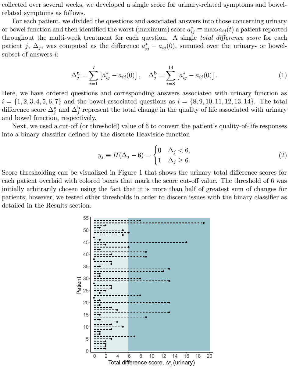
Thanks to advancements in diagnosis and treatment, prostate cancer patients have high long-term survival rates. Currently, an important goal is to preserve quality-of-life during and after treatment. The relationship between the radiation a patient receives and the subsequent side effects he experiences is complex and difficult to model or predict. Here, we use machine learning algorithms and statistical models to explore the connection between radiation treatment and post-treatment gastro-urinary function. Since only a limited number of patient datasets are currently available, we used image flipping and curvature-based interpolation methods to generate more data in order to leverage transfer learning. Using interpolated and augmented data, we trained a convolutional autoencoder network to obtain near-optimal starting points for the weights. A convolutional neural network then analyzed the relationship between patient-reported quality-of-life and radiation. We also used analysis of variance and logistic regression to explore organ sensitivity to radiation and develop dosage thresholds for each organ region. Our findings show no connection between the bladder and quality-of-life scores. However, we found a connection between radiation applied to posterior and anterior rectal regions to changes in quality-of-life. Finally, we estimated radiation therapy dosage thresholds for each organ. Our analysis connects machine learning methods with organ sensitivity, thus providing a framework for informing cancer patient care using patient reported quality-of-life metrics.
Cytotoxic chemotherapy is a common treatment for advanced prostate cancer. These tumors are also known to rely on angiogenesis, i.e., the growth of local microvasculature via chemical signaling produced by the tumor. Thus, several clinical studies have been investigating antiangiogenic therapy for advanced prostate cancer, either as monotherapy or combined with standard cytotoxic protocols. However, the complex genetic alterations promoting prostate cancer growth complicate the selection of the best chemotherapeutic approach for each patient's tumor. Here, we present a mathematical model of prostate cancer growth and chemotherapy that may enable physicians to test and design personalized chemotherapeutic protocols in silico. We use the phase-field method to describe tumor growth, which we assume to be driven by a generic nutrient following reaction-diffusion dynamics. Tumor proliferation and apoptosis (i.e., programmed cell death) can be parameterized with experimentally-determined values. Cytotoxic chemotherapy is included as a term downregulating tumor net proliferation, while antiangiogenic therapy is modeled as a reduction in intratumoral nutrient supply. Another equation couples the tumor phase field with the production of prostate-specific antigen, which is an extensively used prostate cancer biomarker. We prove the well-posedness of our model and we run a series of representative simulations using an isogeometric method to explore untreated tumor growth as well as the effects of cytotoxic chemotherapy and antiangiogenic therapy, both alone and combined. Our simulations show that our model captures the growth morphologies of prostate cancer as well as common outcomes of cytotoxic and antiangiogenic mono and combined therapy. Our model also reproduces the usual temporal trends in tumor volume and prostate-specific antigen evolution observed in previous studies.
Differentiability of the control-to-state map is shown for a three-equation system with general fractional diffusion operators.
abstractclick to expand
In this paper, the authors study the distributed optimal control of a system of three evolutionary equations involving fractional powers of three selfadjoint, monotone, unbounded linear operators having compact resolvents. The system is a generalization of a Cahn-Hilliard type phase field system modeling tumor growth that goes back to Hawkins-Daarud et al. (Int. J. Numer. Math. Biomed. Eng. 28 (2012), 3-24). The aim of the control process, which could be realized by either administering a drug or monitoring the nutrition, is to keep the tumor cell fraction under control while avoiding possible harm for the patient. In contrast to previous studies, in which the occurring unbounded operators governing the diffusional regimes were all given by the Laplacian with zero Neumann boundary conditions, the operators may in our case be different; more generally, we consider systems with fractional powers of the type that were studied in the recent work Adv. Math. Sci. Appl. 28 (2019), 343-375 (see arXiv:1906.10874), by the present authors. In the analysis, the Fr\'echet differentiability of the associated control-to-state operator is shown, by also establishing the existence of solutions to the associated adjoint system, and deriving the first-order necessary conditions of optimality for a cost functional of tracking type.
During development, embryos perform a mesmerizing choreography, which is crucial for the correct shaping, positioning and function of all organs. The cellular properties powering animal morphogenesis have been the focus of much attention. On the other hand, much less consideration has been given to the invisible engine constituted by the intercellular fluid. Cells are immersed in fluid, of which the composition and physical properties have a considerable impact on development. In this review, we revisit recent studies from the perspective of the fluid, focusing on basolateral fluid compartments and taking the early mouse and zebrafish embryos as models. These examples illustrate how the hydration levels of tissues are spatio-temporally controlled and influence embryonic development.
Understanding how the spatial structure of blood vessel networks relates to their function in healthy and abnormal biological tissues could improve diagnosis and treatment for diseases such as cancer. New imaging techniques can generate multiple, high-resolution images of the same tissue region, and show how vessel networks evolve during disease onset and treatment. Such experimental advances have created an exciting opportunity for discovering new links between vessel structure and disease through the development of mathematical tools that can analyse these rich datasets. Here we explain how topological data analysis (TDA) can be used to study vessel network structures. TDA is a growing field in the mathematical and computational sciences, that consists of algorithmic methods for identifying global and multi-scale structures in high-dimensional data sets that may be noisy and incomplete. TDA has identified the effect of ageing on vessel networks in the brain and more recently proposed to study blood flow and stenosis. Here we present preliminary work which shows how TDA of spatial network structure can be used to characterise tumour vasculature.
3D latent vectors from synthetic patient data separate into clusters that may mark subtypes with different treatment responses.
abstractclick to expand
In this paper, we seek a clinically-relevant latent code for representing the spectrum of macular disease. Towards this end, we construct retina-VAE, a variational autoencoder-based model that accepts a patient profile vector (pVec) as input. The pVec components include clinical exam findings and demographic information. We evaluate the model on a subspectrum of the retinal maculopathies, in particular, exudative age-related macular degeneration, central serous chorioretinopathy, and polypoidal choroidal vasculopathy. For these three maculopathies, a database of 3000 6-dimensional pVecs (1000 each) was synthetically generated based on known disease statistics in the literature. The database was then used to train the VAE and generate latent vector representations. We found training performance to be best for a 3-dimensional latent vector architecture compared to 2 or 4 dimensional latents. Additionally, for the 3D latent architecture, we discovered that the resulting latent vectors were strongly clustered spontaneously into one of 14 clusters. Kmeans was then used only to identify members of each cluster and to inspect cluster properties. These clusters suggest underlying disease subtypes which may potentially respond better or worse to particular pharmaceutical treatments such as anti-vascular endothelial growth factor variants. The retina-VAE framework will potentially yield new fundamental insights into the mechanisms and manifestations of disease. And will potentially facilitate the development of personalized pharmaceuticals and gene therapies.
Using formal asymptotic methods we derive a free boundary problem representing one of the simplest mathematical descriptions of the growth and death of a tumour or other biological tissue. The mathematical model takes the form of a closed interface evolving via forced mean curvature flow (together with a `kinetic under-cooling' regularisation) where the forcing depends on the solution of a PDE that holds in the domain enclosed by the interface. We perform linear stability analysis and derive a diffuse-interface approximation of the model. Finite-element discretisations of two closely related models are presented, together with computational results comparing the approximate solutions.
Multi-stage model analysis shows only stem cell self-renewal shapes long-term clonal dominance, while non-stem parameters matter little.
abstractclick to expand
Recent progress in genetic techniques has shed light on the complex co-evolution of malignant cell clones in leukemias. However, several aspects of clonal selection still remain unclear. In this paper, we present a multi-compartmental continuously structured population model of selection dynamics in acute leukemias, which consists of a system of coupled integro-differential equations. Our model can be analysed in a more efficient way than classical models formulated in terms of ordinary differential equations. Exploiting the analytical tractability of this model, we investigate how clonal selection is shaped by the self-renewal fraction and the proliferation rate of leukemic cells at different maturation stages. We integrate analytical results with numerical solutions of a calibrated version of the model based on real patient data. In summary, our mathematical results formalise the biological notion that clonal selection is driven by the self-renewal fraction of leukemic stem cells and the clones that possess the highest value of this parameter are ultimately selected. Moreover, we demonstrate that the self-renewal fraction and the proliferation rate of non-stem cells do not have a substantial impact on clonal selection. Taken together, our results indicate that interclonal variability in the self-renewal fraction of leukemic stem cells provides the necessary substrate for clonal selection to act upon.
It is still not understood how similar genomic sequences have generated diverse and spectacular forms during evolution. The difficulty to bridge phenotypes and genotypes stems from the complexity of multicellular systems, where thousands of genes and cells interact with each other providing developmental non-linearity. To understand how diverse morphologies have evolved, it is essential to find ways to handle such complex systems. Here, we review the fin-to-limb transition as a case study for the evolution of multicellular systems. We first describe the historical perspective of comparative studies between fins and limbs. Second, we introduce our approach that combines mechanistic theory, computational modeling, and in vivo experiments to provide a mechanical explanation for the morphological difference between fish fins and tetrapod limbs. This approach helps resolve a long-standing debate about anatomical homology between the skeletal elements of fins and limbs. We will conclude by proposing that due to the counter-intuitive dynamics of gene interactions, integrative approaches that combine computer modeling, theory and experiments are essential to understand the evolution of multicellular organisms.
Many questions concerning the biophysical and physiological properties of skin are still open. Skin aging, permeability, dermal absorption, hydration and drug transdermal delivery, are few examples of processes with its underlying mechanisms unveiled. In this work we present a first-principles density functional quantum atomistic model for single layer stratum corneum (SC) in order to contribute to unveil the molecular interactions behind the skin properties at this scale. The molecular structure of SC was modeled by an archetype of its hygroscopic proteic portion inside of the corneocytes, the natural moisturizing factor (NMF), coupled to glycerol molecules which represent the lipid fraction of SC. The vibrational spectra was calculated and compared to Fourier-Transform Infrared Absorption spectroscopy (FTIR) experimental data obtained on animal model of SC. We noticed that bands in the fingerprint region (800-1800 cm$^{-1}$) were correctly assigned. Moreover, our calculations revealed the existence of two coupled vibration between hydroxyl group of lipid and NMF methylene (1120 and 1160 cm$^{-1}$), which are of special interest since they probe the lipid-amino acid coupling. The model was also able to predict the shear modulus of dry SC in excellent agreement with the reported value on literature. Others physical/chemical properties could be calculated exploring the chemical accuracy and molecular resolution of our model. Research in dermatology, cosmetology, and biomedical engineering in the specific topics of drug delivery and/or mechanical properties of skin are examples of fields that would potentially take advantage of our approach.
Local cancer invasion of tissue is a complex, multiscale process which plays an essential role in tumour progression. Occurring over many different temporal and spatial scales, the first stage of invasion is the secretion of matrix degrading enzymes (MDEs) by the cancer cells that consequently degrade the surrounding extracellular matrix (ECM). This process is vital for creating space in which the cancer cells can progress and it is driven by the activities of specific matrix metalloproteinases (MMPs). In this paper, we consider the key role of two MMPs by developing further the novel two-part multiscale model introduced in [33] to better relate at micro-scale the two micro-scale activities that were considered there, namely, the micro-dynamics concerning the continuous rearrangement of the naturally oriented ECM fibres within the bulk of the tumour and MDEs proteolytic micro-dynamics that take place in an appropriate cell-scale neighbourhood of the tumour boundary. Focussing primarily on the activities of the membrane-tethered MT1-MMP and the soluble MMP-2 with the fibrous ECM phase, in this work we investigate the MT1-MMP/MMP-2 cascade and its overall effect on tumour progression. To that end, we will propose a new multiscale modelling framework by considering the degradation of the ECM fibres not only to take place at macro-scale in the bulk of the tumour but also explicitly in the micro-scale neighbourhood of the tumour interface as a consequence of the interactions with molecular fluxes of MDEs that exercise their spatial dynamics at the invasive edge of the tumour.
Local cancer cell invasion is a complex process involving many cellular and tissue interactions and is an important prerequisite for metastatic spread, the main cause of cancer related deaths. Occurring over many different temporal and spatial scales, the first stage of local invasion is the secretion of matrix-degrading enzymes (MDEs) and the resulting degradation of the extra-cellular matrix (ECM). This process creates space in which the cells can invade and thus enlarge the tumour. As a tumour increases in malignancy, the cancer cells adopt the ability to mutate into secondary cell subpopulations giving rise to a heterogeneous tumour. This new cell subpopulation often carries higher invasive qualities and permits a quicker spread of the tumour. Building upon the recent multiscale modelling framework for cancer invasion within a fibrous ECM introduced in Shuttleworth and Trucu (2019), in this paper we consider the process of local invasion by a heterotypic tumour consisting of two cancer cell populations mixed with a two-phase ECM. To that end, we address the double feedback link between the tissue-scale cancer dynamics and the cell-scale molecular processes through the development of a two-part modelling framework that crucially incorporates the multiscale dynamic redistribution of oriented fibres occurring within a two-phase extra-cellular matrix and combines this with the multiscale leading edge dynamics exploring key matrix-degrading enzymes molecular processes along the tumour interface that drive the movement of the cancer boundary. The modelling framework will be accompanied by computational results that explore the effects of the underlying fibre network on the overall pattern of cancer invasion.