RadiomicNet: A Hybrid Radiomics-Guided Lightweight Architecture for Interpretable Medical Image Segmentation
Pith reviewed 2026-07-03 15:59 UTC · model grok-4.3
The pith
Integrating GLCM and LBP radiomics features via an attention gate yields higher Dice scores in a lightweight segmentation network.
A machine-rendered reading of the paper's core claim, the machinery that carries it, and where it could break.
Core claim
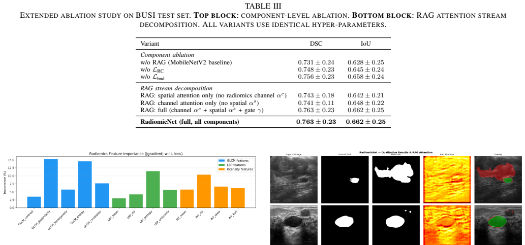
By feeding Gray-Level Co-occurrence Matrix and Local Binary Pattern features into a Radiomics Attention Gate that modulates skip-connection attention inside a MobileNetV2-based encoder-decoder and adding a Radiomics Consistency Loss that aligns texture complexity with prediction uncertainty, RadiomicNet reaches Dice Similarity Coefficients of 0.763 on BUSI and 0.854 on Kvasir-SEG, exceeds U-KAN performance, uses only 3.27 million parameters, lowers Expected Calibration Error from 0.142 to 0.118, and supplies ante-hoc interpretability through feature-importance rankings of GLCM dissimilarity, energy, and LBP entropy.
What carries the argument
The Radiomics Attention Gate (RAG), which uses GLCM and LBP features to modulate skip-connection attention.
If this is right
- Segmentation accuracy can be maintained or improved while shrinking parameter count by nearly an order of magnitude relative to standard U-Net.
- Direct incorporation of domain texture descriptors can reduce expected calibration error without post-training adjustments.
- Gradient-based ranking of individual radiomics features supplies clinically readable explanations for each segmentation output.
- The same two-stream pattern can be applied across different medical imaging tasks that already use GLCM or LBP descriptors.
Where Pith is reading between the lines
- The approach may lessen reliance on separate post-hoc explanation tools in clinical deployment.
- Testing the same radiomics gate on CT or MRI volumes could reveal whether the parameter savings and calibration benefit hold beyond ultrasound and endoscopy.
- The consistency loss term might be ported to other uncertainty-sensitive tasks such as lesion detection or registration.
Load-bearing premise
The reported gains in accuracy, calibration, and interpretability arise specifically from the addition of the radiomics attention gate and consistency loss rather than from other unstated architectural choices or dataset-specific tuning.
What would settle it
Training the identical MobileNetV2 backbone on the same BUSI and Kvasir-SEG splits without the Radiomics Attention Gate or the consistency loss and checking whether Dice scores and ECE fall to or below the U-KAN and U-Net levels.
Figures


read the original abstract
Deep learning has achieved remarkable performance in medical image segmentation, yet it suffers from critical limitations: mathematical intractability, substantial parameter requirements, and lack of clinical interpretability. We propose RadiomicNet, a novel two-stream hybrid architecture that enhances standard deep learning by integrating handcrafted radiomics features directly into the segmentation learning process. The key contribution is the Radiomics Attention Gate (RAG), which leverages Gray-Level Co-occurrence Matrix (GLCM) and Local Binary Pattern (LBP) features to modulate skip-connection attention in a lightweight MobileNetV2-based encoder-decoder, providing ante-hoc interpretability without post-hoc approximations. A novel Radiomics Consistency Loss further enforces alignment between texture complexity and prediction uncertainty, reducing Expected Calibration Error (ECE) from 0.142 to 0.118. RadiomicNet achieves a Dice Similarity Coefficient (DSC) of 0.763 +/- 0.231 on the Breast Ultrasound Images (BUSI) dataset and 0.854 +/- 0.112 on Kvasir-SEG, outperforming U-KAN by 1.2% and 1.8%, respectively (p < 0.05, Wilcoxon signed-rank test), with only 3.27M parameters, 9.5x fewer than standard U-Net and 4.3x fewer than U-KAN. Gradient-based feature importance analysis reveals that GLCM dissimilarity (15.24%), GLCM energy (14.56%), and LBP entropy (11.49%) are the dominant radiomics cues, providing clinically meaningful explanations for segmentation decisions. The proposed approach demonstrates that compact, interpretable models grounded in domain knowledge can deliver state-of-the-art segmentation performance with substantially reduced computational overhead.
Editorial analysis
A structured set of objections, weighed in public.
Referee Report
Summary. The paper introduces RadiomicNet, a two-stream hybrid model combining a MobileNetV2 encoder-decoder with radiomics features (GLCM, LBP) via the Radiomics Attention Gate (RAG) for skip connections, and a Radiomics Consistency Loss to reduce calibration error. It reports superior DSC on BUSI (0.763±0.231) and Kvasir-SEG (0.854±0.112) datasets compared to U-KAN, with fewer parameters and improved interpretability through feature importance analysis.
Significance. Should the gains prove attributable to the proposed RAG and consistency mechanisms rather than unablated factors, this would represent a meaningful step toward domain-knowledge-infused lightweight models in medical segmentation. The emphasis on ante-hoc interpretability and efficiency (3.27M parameters) addresses key barriers to clinical translation. The gradient-based analysis of radiomics feature importance provides a concrete example of how such models can offer clinically relevant explanations.
major comments (2)
- [Abstract] Abstract: The DSC improvements (1.2% and 1.8% over U-KAN) and ECE reduction (0.142 to 0.118) are presented without ablation experiments that isolate the Radiomics Attention Gate (e.g., by replacing it with standard skip connections) or the consistency loss while holding the MobileNetV2 backbone, optimizer, augmentation, and data splits fixed. This omission leaves the causal contribution of the novel components unverified.
- [Methods] Methods (Radiomics Consistency Loss): The loss is described as enforcing alignment between texture complexity and prediction uncertainty, but no explicit equation or implementation detail is provided for how texture complexity is computed from GLCM/LBP or how the term is weighted in the total loss. Without this, it is unclear whether the ECE improvement is independent or tied to dataset-specific hyperparameter tuning.
minor comments (2)
- A table listing parameter counts, FLOPs, and inference times for all compared models (U-Net, U-KAN, RadiomicNet) would clarify the efficiency claims.
- The number of runs, cross-validation folds, or paired samples underlying the Wilcoxon signed-rank test and the reported standard deviations should be stated explicitly.
Simulated Author's Rebuttal
We thank the referee for their constructive comments, which highlight important aspects for strengthening the manuscript. We address each major comment below with clarifications and commitments to revisions where the points are valid.
read point-by-point responses
-
Referee: [Abstract] Abstract: The DSC improvements (1.2% and 1.8% over U-KAN) and ECE reduction (0.142 to 0.118) are presented without ablation experiments that isolate the Radiomics Attention Gate (e.g., by replacing it with standard skip connections) or the consistency loss while holding the MobileNetV2 backbone, optimizer, augmentation, and data splits fixed. This omission leaves the causal contribution of the novel components unverified.
Authors: We agree that the manuscript would benefit from explicit ablation studies to isolate the contributions of the Radiomics Attention Gate (RAG) and the Radiomics Consistency Loss. In the revised version, we will add these experiments, including variants where RAG is replaced by standard skip connections and where the consistency loss is ablated, while fixing the MobileNetV2 backbone, optimizer, augmentations, and data splits. This will directly verify the causal impact of the proposed components on DSC and ECE. revision: yes
-
Referee: [Methods] Methods (Radiomics Consistency Loss): The loss is described as enforcing alignment between texture complexity and prediction uncertainty, but no explicit equation or implementation detail is provided for how texture complexity is computed from GLCM/LBP or how the term is weighted in the total loss. Without this, it is unclear whether the ECE improvement is independent or tied to dataset-specific hyperparameter tuning.
Authors: We acknowledge the lack of explicit implementation details for the Radiomics Consistency Loss. In the revised manuscript, we will include the full mathematical formulation of the loss, specifying how texture complexity is computed from GLCM (e.g., via contrast, dissimilarity, or energy) and LBP (e.g., via entropy or uniformity) features, along with the weighting coefficient in the total loss. We will also report the hyperparameter values used and any relevant sensitivity checks to clarify independence from dataset-specific tuning. revision: yes
Circularity Check
No circularity: empirical architecture proposal with reported metrics
full rationale
The paper proposes RadiomicNet as a hybrid architecture incorporating GLCM/LBP features via RAG and a consistency loss, then reports end-to-end DSC and ECE numbers on BUSI and Kvasir-SEG. No derivation chain, uniqueness theorem, or first-principles prediction is claimed; performance figures are presented as experimental outcomes rather than quantities forced by construction from fitted inputs or self-citations. The consistency loss is introduced as a novel term whose effect is measured externally on held-out metrics, satisfying the criterion for independent content.
Axiom & Free-Parameter Ledger
invented entities (1)
-
Radiomics Attention Gate (RAG)
no independent evidence
Reference graph
Works this paper leans on
-
[1]
U-Net: Convolutional net- works for biomedical image segmentation,
O. Ronneberger, P. Fischer, and T. Brox, “U-Net: Convolutional net- works for biomedical image segmentation,” inProc. MICCAI, 2015, pp. 234–241
work page 2015
-
[2]
UNet++: A nested U-Net architecture for medical image segmentation,
Z. Zhou, M. M. R. Siddiquee, N. Tajbakhsh, and J. Liang, “UNet++: A nested U-Net architecture for medical image segmentation,” inDeep Learning in Medical Image Analysis, 2018, pp. 3–11
work page 2018
-
[3]
J. Chenet al., “TransUNet: Rethinking the U-Net architecture design for medical image segmentation through the lens of transformers,”Medical Image Analysis, vol. 97, 2024
work page 2024
-
[4]
U-KAN makes strong backbone for medical image segmentation and generation,
C. Li, X. Liu, W. Li, C. Wang, H. Liu, and Y . Yuan, “U-KAN makes strong backbone for medical image segmentation and generation,” in Proc. AAAI, 2025, pp. 4652–4660
work page 2025
-
[5]
IKANet: A Kolmogorov-Arnold network for interpretable medical image segmenta- tion,
M. Chen, J. Zhang, L. Jin, X. Xuan, and Y . Cheng, “IKANet: A Kolmogorov-Arnold network for interpretable medical image segmenta- tion,” inProc. PRCV, 2025
work page 2025
-
[6]
KAN: Kolmogorov-Arnold networks,
Z. Liuet al., “KAN: Kolmogorov-Arnold networks,” inProc. ICLR, 2025
work page 2025
-
[7]
Radiomics: Extracting more information from medical images using advanced feature analysis,
P. Lambinet al., “Radiomics: Extracting more information from medical images using advanced feature analysis,”Eur. J. Cancer, vol. 48, no. 4, pp. 441–446, 2012
work page 2012
-
[8]
Radiomics: Images are more than pictures, they are data,
R. J. Gillies, P. E. Kinahan, and H. Hricak, “Radiomics: Images are more than pictures, they are data,”Radiology, vol. 278, no. 2, pp. 563–577, 2016
work page 2016
-
[9]
Focus on texture: Rethinking pre-training in masked autoencoders for medical image classification,
S. Basuet al., “Focus on texture: Rethinking pre-training in masked autoencoders for medical image classification,” inProc. MICCAI, 2025
work page 2025
-
[10]
B. Baidya Kayalet al., “Advances in medical image segmentation: A comprehensive review of traditional, deep learning and hybrid ap- proaches,”Bioengineering, vol. 11, no. 10, 2024
work page 2024
-
[11]
Dataset of breast ultrasound images,
W. Al-Dhabyani, M. Gomaa, H. Khaled, and A. Fahmy, “Dataset of breast ultrasound images,”Data in Brief, vol. 28, 2020
work page 2020
-
[12]
Kvasir-SEG: A segmented polyp dataset,
D. Jhaet al., “Kvasir-SEG: A segmented polyp dataset,” inProc. MMM, 2020, pp. 451–462
work page 2020
-
[13]
MobileNetV2: Inverted residuals and linear bottlenecks,
M. Sandler, A. Howard, M. Zhu, A. Zhmoginov, and L.-C. Chen, “MobileNetV2: Inverted residuals and linear bottlenecks,” inProc. CVPR, 2018
work page 2018
-
[14]
The second workshop on learning beyond deep learning (LBDL II),
C.-C. J. Kuo and L. Guan, “The second workshop on learning beyond deep learning (LBDL II),” inProc. IEEE ICIP Workshop, 2026
work page 2026
discussion (0)
Sign in with ORCID, Apple, or X to comment. Anyone can read and Pith papers without signing in.