X-Splat: Gaussian Splatting for 3D CBCT Generation from Single Panoramic Radiograph
Pith reviewed 2026-07-03 15:33 UTC · model grok-4.3
The pith
X-Splat recovers sharp 3D dental structures like individual teeth and the mandibular canal from a single panoramic radiograph by initializing anisotropic Gaussians along the acquisition paths.
A machine-rendered reading of the paper's core claim, the machinery that carries it, and where it could break.
Core claim
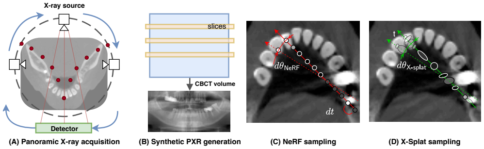
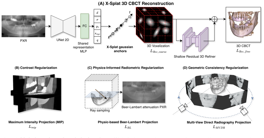
X-Splat is the first Gaussian Splatting framework for CBCT generation from single PXR. It uses the known panoramic acquisition geometry as a scaffold by initializing anisotropic Gaussian primitives along the X-ray paths that formed the input image, then adjusts them in a single feed-forward pass constrained by Beer-Lambert reprojection and multi-view radiographic supervision. A lightweight residual refiner adds dataset-level anatomical priors. Trained on synthetic PXR-CBCT pairs, the method recovers individual teeth, cortical boundaries, alveolar structure, and the mandibular canal that prior NeRF- and GAN-based baselines fail to reconstruct.
What carries the argument
Anisotropic Gaussian primitives initialized along known panoramic X-ray paths and adjusted under Beer-Lambert reprojection and multi-view supervision.
If this is right
- Individual teeth and cortical boundaries become visible in the output volume.
- Alveolar structure including the mandibular canal is recovered where prior methods produce none.
- Geometry-driven supervision from the panoramic paths reduces anatomically inconsistent hallucinations.
- Training on synthetic PXR-CBCT pairs enables direct volumetric supervision without real paired scans.
- Segmentation-based metrics provide the first quantitative evaluation of maxillofacial anatomy recovery from single PXR.
Where Pith is reading between the lines
- The same path-initialization idea could be tested on other single-view radiographic modalities such as chest X-rays to recover 3D structure.
- If the Gaussian representation generalizes, it might allow on-the-fly 3D dental models inside existing panoramic X-ray machines.
- Direct comparison of radiation dose and diagnostic accuracy against standard CBCT protocols would quantify the practical reduction in patient exposure.
- The method's feed-forward nature suggests it could be embedded in real-time clinical software once validated on diverse patient populations.
Load-bearing premise
Initializing and refining Gaussian primitives along the known X-ray paths supplies enough geometric constraint to resolve the missing depth information without hallucinations.
What would settle it
Side-by-side comparison of X-Splat output against real CBCT scans of the same patients, checking whether the reconstructed mandibular canal and tooth roots align in position and shape.
Figures


read the original abstract
Generating a 3D dental volume from a single panoramic radiograph (PXR) could provide a low-radiation alternative to Cone-Beam Computed Tomography (CBCT), but the problem is highly underdetermined: panoramic acquisition integrates 3D attenuation along curved X-ray paths into a 2D image, leaving depth-resolved anatomy unobserved. Existing implicit and generative approaches often produce oversmoothed geometry or anatomically inconsistent hallucinations, lacking geometry-driven supervision and relying on smooth representations unable to precisely localize sharp anatomical boundaries. We propose X-Splat, the first Gaussian Splatting framework for generating CBCT-like 3D dental volumes from a single PXR. X-Splat uses the known panoramic acquisition geometry as a generation scaffold: learnable anisotropic Gaussian primitives are initialized along the X-ray paths that formed the input image and adjusted in a single feed-forward pass, constrained by Beer-Lambert reprojection and multi-view radiographic training supervision. A lightweight residual refiner adds dataset-level anatomical priors without overriding the geometry already resolved by the Gaussians. We train on synthetic PXR-CBCT pairs, enabling direct volumetric supervision without paired real scans. We further introduce segmentation-based geometry-aware metrics, providing the first evaluation of PXR-based generation over maxillofacial anatomy. X-Splat outperforms NeRF- and GAN-based baselines, recovering individual teeth, cortical boundaries, and alveolar structure, including the mandibular canal which prior methods fail to reconstruct. Code will be available at https://github.com/tomek1911/X-Splat
Editorial analysis
A structured set of objections, weighed in public.
Referee Report
Summary. The paper proposes X-Splat, the first Gaussian Splatting framework to generate 3D CBCT-like dental volumes from a single panoramic radiograph (PXR). It initializes learnable anisotropic Gaussian primitives along known X-ray paths from the panoramic acquisition geometry, adjusts them in one feed-forward pass under Beer-Lambert reprojection and multi-view radiographic losses, adds a lightweight residual refiner for anatomical priors, and trains exclusively on synthetic PXR-CBCT pairs. The work claims to outperform NeRF- and GAN-based baselines while recovering fine structures (individual teeth, cortical boundaries, alveolar bone, mandibular canal) that prior methods miss, and introduces segmentation-based geometry-aware metrics for evaluation.
Significance. If the quantitative results and real-data generalization hold, the geometry-driven Gaussian approach could meaningfully advance low-radiation 3D dental imaging by reducing hallucinations common in implicit or generative methods. The explicit use of acquisition geometry as a scaffold, the planned code release, and the new segmentation-based metrics constitute concrete strengths that would support reproducibility and standardized evaluation in this domain.
major comments (2)
- [Abstract] Abstract: the central claim that X-Splat 'outperforms NeRF- and GAN-based baselines' and recovers specific fine structures (teeth, cortical boundaries, alveolar structure, mandibular canal) is presented without any quantitative metrics, error bars, ablation tables, or figure references, leaving the primary performance assertion unsupported in the summary of results.
- [Method] Method description (abstract and §3): the assertion that a single feed-forward adjustment of Gaussians initialized along panoramic paths, constrained only by Beer-Lambert reprojection plus multi-view losses, suffices to resolve depth ambiguities without hallucinations is load-bearing for the central claim yet lacks supporting analysis of residual depth errors or domain-shift behavior on real radiographs.
minor comments (2)
- [Abstract] The statement that code 'will be available' should be updated with a permanent link or DOI once released to fulfill the reproducibility commitment.
- [Experiments] Clarify in the evaluation section how the synthetic PXR-CBCT pairs were generated and whether any quantitative measure of their realism relative to real anatomy is provided.
Simulated Author's Rebuttal
We thank the referee for the constructive feedback and for recognizing the potential of the geometry-driven Gaussian approach. We address each major comment below and indicate planned revisions.
read point-by-point responses
-
Referee: [Abstract] Abstract: the central claim that X-Splat 'outperforms NeRF- and GAN-based baselines' and recovers specific fine structures (teeth, cortical boundaries, alveolar structure, mandibular canal) is presented without any quantitative metrics, error bars, ablation tables, or figure references, leaving the primary performance assertion unsupported in the summary of results.
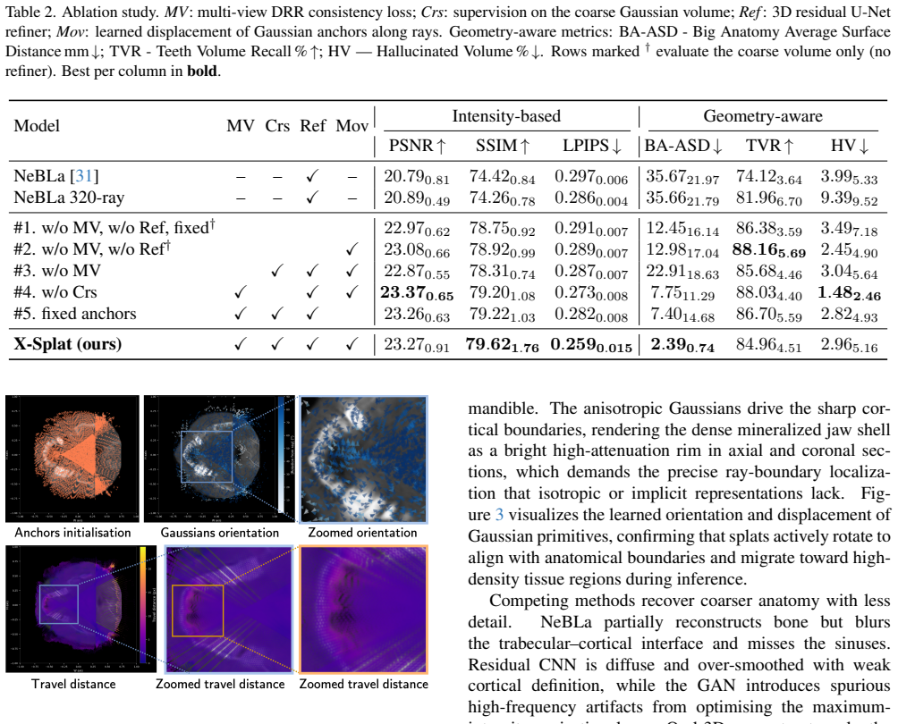
Authors: We agree that the abstract would benefit from tighter linkage to the supporting evidence. The body of the manuscript contains the quantitative results (Table 1), ablation studies (Table 2), and figures (Figs. 3–5) that substantiate the claims. We will revise the abstract to include one or two key metric values and explicit references to the relevant tables and figures. revision: yes
-
Referee: [Method] Method description (abstract and §3): the assertion that a single feed-forward adjustment of Gaussians initialized along panoramic paths, constrained only by Beer-Lambert reprojection plus multi-view losses, suffices to resolve depth ambiguities without hallucinations is load-bearing for the central claim yet lacks supporting analysis of residual depth errors or domain-shift behavior on real radiographs.
Authors: The panoramic-path initialization supplies an explicit geometric scaffold that directly constrains depth; the quantitative superiority over baselines that lack this prior (reported in §4) serves as indirect evidence that depth ambiguities are reduced. We acknowledge, however, that the manuscript does not include an explicit residual-depth-error analysis or systematic domain-shift experiments on real radiographs. We will add a limitations paragraph discussing these points and, where space permits, qualitative real-data examples already present in the supplementary material. revision: partial
Circularity Check
No circularity: method grounded in external physical law and known geometry
full rationale
The paper's core pipeline initializes anisotropic Gaussians along known panoramic X-ray paths and optimizes them under the independent Beer-Lambert attenuation law plus multi-view radiographic losses; these constraints are external physical and geometric facts, not quantities defined in terms of the output volume. Training occurs on synthetic PXR-CBCT pairs generated from an external forward model, enabling direct volumetric supervision without any equation reducing the predicted CBCT to a fitted parameter or self-citation. No self-definitional loops, fitted-input predictions, or load-bearing self-citations appear in the described derivation. The approach therefore remains self-contained against external benchmarks.
Axiom & Free-Parameter Ledger
free parameters (1)
- Gaussian primitive parameters (position, anisotropy, opacity, color)
axioms (2)
- standard math Beer-Lambert law governs X-ray attenuation along each ray path
- domain assumption Synthetic PXR-CBCT pairs provide unbiased volumetric ground truth
Reference graph
Works this paper leans on
-
[1]
Ander Biguri, Tomoyuki Sadakane, Reuben Lindroos, Yi Liu, Malena Sabat´e Landman, Yi Du, Manasavee Lohvithee, Stefanie Kaser, Sepideh Hatamikia, Robert Bryll, et al. Ti- gre v3: Efficient and easy to use iterative computed tomo- graphic reconstruction toolbox for real datasets.Engineering Research Express, 7(1):015011, 2025. 12
work page 2025
-
[2]
Federico Bolelli, Luca Lumetti, Shankeeth Vinayahalingam, Mattia Di Bartolomeo, Arrigo Pellacani, Kevin Marchesini, Niels Van Nistelrooij, Pieter Van Lierop, Tong Xi, Yusheng Liu, et al. Segmenting the inferior alveolar canal in cbcts volumes: the toothfairy challenge.IEEE Transactions on Medical Imaging, 44(4):1890–1906, 2024. 2
work page 1906
-
[3]
Segmenting Max- illofacial Structures in CBCT V olume
Federico Bolelli, Kevin Marchesini, Niels van Nistelrooij, Luca Lumetti, Vittorio Pipoli, Elisa Ficarra, Shankeeth Vinayahalingam, and Costantino Grana. Segmenting Max- illofacial Structures in CBCT V olume. InIEEE/CVF Confer- ence on Computer Vision and Pattern Recognition (CVPR), pages 1–10. IEEE, 2025. 5, 13, 14
work page 2025
-
[4]
Segmenting max- illofacial structures in cbct volumes
Federico Bolelli, Kevin Marchesini, Niels Van Nistelrooij, Luca Lumetti, Vittorio Pipoli, Elisa Ficarra, Shankeeth Vinayahalingam, and Costantino Grana. Segmenting max- illofacial structures in cbct volumes. InProceedings of the Computer Vision and Pattern Recognition Conference, pages 5238–5248, 2025. 2, 6, 17
work page 2025
-
[5]
Federico Bolelli, Luca Lumetti, Niels van Nistelrooij, Shankeeth Vinayahalingam, Mattia Di Bartolomeo, Kevin Marchesini, Arrigo Pellacani, Ettore Candeloro, Gabriele Rosati, Tong Xi, et al. Multi-structure segmentation in cbct volumes: The toothfairy2 challenge.Medical Image Analy- sis, page 104095, 2026. 2, 16
work page 2026
-
[6]
Radiative gaussian splatting for efficient x-ray novel view synthesis
Yuanhao Cai, Yixun Liang, Jiahao Wang, Angtian Wang, Yu- lun Zhang, Xiaokang Yang, Zongwei Zhou, and Alan Yuille. Radiative gaussian splatting for efficient x-ray novel view synthesis. InEuropean Conference on Computer Vision, pages 283–299. Springer, 2024. 2, 12
work page 2024
-
[7]
Structure-aware sparse-view x-ray 3d re- construction
Yuanhao Cai, Jiahao Wang, Alan Yuille, Zongwei Zhou, and Angtian Wang. Structure-aware sparse-view x-ray 3d re- construction. InProceedings of the IEEE/CVF conference on computer vision and pattern recognition, pages 11174– 11183, 2024. 12
work page 2024
-
[8]
3d-r2n2: A unified approach for single and multi-view 3d object reconstruction
Christopher B Choy, Danfei Xu, JunYoung Gwak, Kevin Chen, and Silvio Savarese. 3d-r2n2: A unified approach for single and multi-view 3d object reconstruction. InEuropean conference on computer vision, pages 628–644. Springer,
-
[9]
Generative adversarial networks.Commu- nications of the ACM, 63(11):139–144, 2020
Ian Goodfellow, Jean Pouget-Abadie, Mehdi Mirza, Bing Xu, David Warde-Farley, Sherjil Ozair, Aaron Courville, and Yoshua Bengio. Generative adversarial networks.Commu- nications of the ACM, 63(11):139–144, 2020. 6, 17
work page 2020
-
[10]
Generative adversarial nets.Advances in neural information processing systems, 27, 2014
Ian J Goodfellow, Jean Pouget-Abadie, Mehdi Mirza, Bing Xu, David Warde-Farley, Sherjil Ozair, Aaron Courville, and Yoshua Bengio. Generative adversarial nets.Advances in neural information processing systems, 27, 2014. 1, 6, 12
work page 2014
-
[11]
Single-image tomography: 3d volumes from 2d cranial x-rays
Phlipp Henzler, V olker Rasche, Timo Ropinski, and Tobias Ritschel. Single-image tomography: 3d volumes from 2d cranial x-rays. InComputer graphics forum, pages 377–388. Wiley Online Library, 2018. 6, 12, 17
work page 2018
-
[12]
Jonathan Ho, Ajay Jain, and Pieter Abbeel. Denoising dif- fusion probabilistic models.Advances in neural information processing systems, 33:6840–6851, 2020. 1
work page 2020
-
[13]
Lrm: Large reconstruction model for single image to 3d
Yicong Hong, Kai Zhang, Jiuxiang Gu, Sai Bi, Yang Zhou, Difan Liu, Feng Liu, Kalyan Sunkavalli, Trung Bui, and Hao Tan. Lrm: Large reconstruction model for single image to 3d. InInternational Conference on Learning Representa- tions, pages 50678–50702, 2024. 12
work page 2024
-
[14]
3d gaussian splatting for real-time radiance field rendering.ACM Trans
Bernhard Kerbl, Georgios Kopanas, Thomas Leimk ¨uhler, George Drettakis, et al. 3d gaussian splatting for real-time radiance field rendering.ACM Trans. Graph., 42(4):139–1,
-
[15]
3dpx: Progressive 2d-to-3d oral image reconstruction with hybrid mlp-cnn networks
Xiaoshuang Li, Mingyuan Meng, Zimo Huang, Lei Bi, Ed- uardo Delamare, Dagan Feng, Bin Sheng, and Jinman Kim. 3dpx: Progressive 2d-to-3d oral image reconstruction with hybrid mlp-cnn networks. InInternational Conference on Medical Image Computing and Computer-Assisted Interven- tion, pages 25–34. Springer, 2024. 1, 12
work page 2024
-
[16]
Yingtai Li, Xueming Fu, Han Li, Shang Zhao, Ruiyang Jin, and S Kevin Zhou. 3dgr-ct: Sparse-view ct reconstruction with a 3d gaussian representation.Medical Image Analysis, 103:103585, 2025. 12
work page 2025
-
[17]
X2teeth: 3d teeth reconstruction from a single panoramic radiograph
Yuan Liang, Weinan Song, Jiawei Yang, Liang Qiu, Kun Wang, and Lei He. X2teeth: 3d teeth reconstruction from a single panoramic radiograph. InInternational Conference on Medical Image Computing and Computer-Assisted Inter- vention, pages 400–409. Springer, 2020. 1, 12
work page 2020
-
[18]
Tianyu Lin, Xinran Li, Chuntung Zhuang, Qi Chen, Yuan- hao Cai, Kai Ding, Alan Yuille, and Zongwei Zhou. Are pixel-wise metrics reliable for computerized tomography re- construction?Advances in Neural Information Processing Systems, 38:33662–33708, 2026. 2, 12
work page 2026
-
[19]
Zhentao Liu, Yu Fang, Changjian Li, Han Wu, Yuan Liu, Dinggang Shen, and Zhiming Cui. Geometry-aware atten- uation learning for sparse-view cbct reconstruction.IEEE Transactions on Medical Imaging, 44(2):1083–1097, 2024. 1, 12
work page 2024
-
[20]
Zhentao Liu, Huangxuan Zhao, Wenhui Qin, Zhenghong Zhou, Xinggang Wang, Wenping Wang, Xiaochun Lai, Dinggang Shen, and Zhiming Cui. 3d vessel reconstruction from sparse-view dynamic dsa images via vessel probability guided attenuation learning.Medical Image Analysis, page 104088, 2026. 12 9
work page 2026
-
[21]
Wonder3d: Sin- gle image to 3d using cross-domain diffusion
Xiaoxiao Long, Yuan-Chen Guo, Cheng Lin, Yuan Liu, Zhiyang Dou, Lingjie Liu, Yuexin Ma, Song-Hai Zhang, Marc Habermann, Christian Theobalt, et al. Wonder3d: Sin- gle image to 3d using cross-domain diffusion. InProceed- ings of the IEEE/CVF conference on computer vision and pattern recognition, pages 9970–9980, 2024. 12
work page 2024
-
[22]
Luca Lumetti, Vittorio Pipoli, Federico Bolelli, Elisa Ficarra, and Costantino Grana. Enhancing Patch-Based Learning for the Segmentation of the Mandibular Canal.IEEE Access, pages 1–12, 2024. 5, 13, 14
work page 2024
-
[23]
Toothfairy3: Scaling cbct maxillofacial segmentation to 77 classes with u-mamba2
Luca Lumetti, Zhi Qin Tan, Lorenzo Borghi, Owen Addison, Yupeng Li, Gabriele Rosati, Niels van Nistelrooij, Shankeeth Vinayahalingam, Costantino Grana, and Federico Bolelli. Toothfairy3: Scaling cbct maxillofacial segmentation to 77 classes with u-mamba2. InMedical Image Computing and Computer Assisted Intervention – MICCAI 2026, 2026. 5, 13, 14
work page 2026
-
[24]
Px2tooth: Reconstructing the 3d point cloud teeth from a single panoramic x-ray
Wen Ma, Huikai Wu, Zikai Xiao, Yang Feng, Jian Wu, and Zuozhu Liu. Px2tooth: Reconstructing the 3d point cloud teeth from a single panoramic x-ray. InInternational Confer- ence on Medical Image Computing and Computer-Assisted Intervention, pages 411–421. Springer, 2024. 1, 12
work page 2024
-
[25]
David MacDonald, Sharifa Alebrahim, Edwin Yen, and Jolanta Aleksejuniene. Cone-beam computed tomographic reconstructions in the evaluation of maxillary impacted ca- nines.Imaging science in dentistry, 53(2):145, 2023. 1
work page 2023
-
[26]
Lanzhuju Mei, Yu Fang, Yue Zhao, Xiang Sean Zhou, Min Zhu, Zhiming Cui, and Dinggang Shen. Dtr-net: dual-space 3d tooth model reconstruction from panoramic x-ray im- ages.IEEE Transactions on Medical Imaging, 43(1):517– 528, 2023. 12
work page 2023
-
[27]
Ben Mildenhall, Pratul P Srinivasan, Matthew Tancik, Jonathan T Barron, Ravi Ramamoorthi, and Ren Ng. Nerf: Representing scenes as neural radiance fields for view syn- thesis.Communications of the ACM, 65(1):99–106, 2021. 1, 5, 13
work page 2021
-
[28]
Sumeet Minhas, Tai-Hsien Wu, Do-Gyoon Kim, Si Chen, Yi-Chu Wu, and Ching-Chang Ko. Artificial intelligence for 3d reconstruction from 2d panoramic x-rays to assess maxil- lary impacted canines.Diagnostics, 14(2):196, 2024. 1
work page 2024
-
[29]
Society for Industrial and Applied Mathematics, Philadelphia, PA, 2001
Frank Natterer.The Mathematics of Computerized Tomog- raphy. Society for Industrial and Applied Mathematics, Philadelphia, PA, 2001. 5
work page 2001
-
[30]
3d teeth reconstruction from panoramic radiographs using neural implicit functions
Sihwa Park, Seongjun Kim, In-Seok Song, and Seung Jun Baek. 3d teeth reconstruction from panoramic radiographs using neural implicit functions. InInternational Conference on Medical Image Computing and Computer-Assisted Inter- vention, pages 376–386. Springer, 2023. 1, 12
work page 2023
-
[31]
Nebla: Neural beer-lambert for 3d reconstruction of oral structures from panoramic radiographs
Sihwa Park, Seongjun Kim, Doeyoung Kwon, Yohan Jang, In-Seok Song, and Seung Jun Baek. Nebla: Neural beer-lambert for 3d reconstruction of oral structures from panoramic radiographs. InProceedings of the AAAI Con- ference on Artificial Intelligence, pages 4433–4441, 2024. 1, 2, 3, 4, 5, 6, 7, 12, 13, 14, 16, 17
work page 2024
-
[32]
Ruben Pauwels, Jilke Beinsberger, Bruno Collaert, Chrysoula Theodorakou, Jessica Rogers, Anne Walker, Lesley Cockmartin, Hilde Bosmans, Reinhilde Jacobs, Ria Bogaerts, et al. Effective dose range for dental cone beam computed tomography scanners.European journal of radiology, 81(2):267–271, 2012. 1
work page 2012
-
[33]
Pawel Tomasz Pieta, Rasmus Juul Pedersen, Sina Borgi, Jakob Sauer Jørgensen, Jens Wenzel Andreasen, and Ve- drana Andersen Dahl. Fact-gs: Fast and scalable ct reconstruction with gaussian splatting.arXiv preprint arXiv:2604.01844, 2026. 12
-
[34]
Panoramic radiography in den- tistry
Ingrid Rozylo-Kalinowska. Panoramic radiography in den- tistry. InImaging techniques in dental radiology: acquisi- tion, anatomic analysis and interpretation of radiographic images, pages 43–56. Springer, 2020. 1
work page 2020
-
[35]
Neat: Neural adaptive tomography
Darius R ¨uckert, Yuanhao Wang, Rui Li, Ramzi Idoughi, and Wolfgang Heidrich. Neat: Neural adaptive tomography. ACM Transactions on Graphics (TOG), 41(4):1–13, 2022. 12
work page 2022
-
[36]
William C. Scarfe and Allan G. Farman. What is cone-beam CT and how does it work?Dental Clinics of North America, 52(4):707–730, 2008. 5
work page 2008
-
[37]
RKW Schulze and NA Drage. Cone-beam computed tomog- raphy and its applications in dental and maxillofacial radiol- ogy.Clinical radiology, 75(9):647–657, 2020. 1
work page 2020
-
[38]
Oral-3d: Reconstructing the 3d structure of oral cavity from panoramic x-ray
Weinan Song, Yuan Liang, Jiawei Yang, Kun Wang, and Lei He. Oral-3d: Reconstructing the 3d structure of oral cavity from panoramic x-ray. InProceedings of the AAAI confer- ence on artificial intelligence, pages 566–573, 2021. 1, 2, 6, 12, 16, 17
work page 2021
-
[39]
Let me decode you: Decoder conditioning with tabular data
Tomasz Szczepa ´nski, Michal K Grzeszczyk, Szymon Płotka, Arleta Adamowicz, Piotr Fudalej, Przemysław Ko- rzeniowski, Tomasz Trzci´nski, and Arkadiusz Sitek. Let me decode you: Decoder conditioning with tabular data. InIn- ternational Conference on Medical Image Computing and Computer-Assisted Intervention, pages 228–238. Springer,
-
[40]
Gepar3d: Geometry prior-assisted learning for 3d tooth segmentation
Tomasz Szczepa ´nski, Szymon Płotka, Michal K Grzeszczyk, Arleta Adamowicz, Piotr Fudalej, Przemysław Ko- rzeniowski, Tomasz Trzci ´nski, and Arkadiusz Sitek. Gepar3d: Geometry prior-assisted learning for 3d tooth segmentation. InInternational Conference on Medical Image Computing and Computer-Assisted Intervention, pages 218–228. Springer, 2025. 2, 13
work page 2025
-
[41]
Springer Nature Switzerland, 2026
Tomasz Szczepa ´nski and Szymon Płotka.Morphology- Driven Deep Watershed Transform for 3D Tooth Segmenta- tion, page 159–167. Springer Nature Switzerland, 2026. 2, 13
work page 2026
-
[42]
Haoshen Wang, Zhentao Liu, Kaicong Sun, Xiaodong Wang, Dinggang Shen, and Zhiming Cui. 3d meddiffusion: A 3d medical latent diffusion model for controllable and high- quality medical image generation.IEEE Transactions on Medical Imaging, 2025. 1
work page 2025
-
[43]
Jiale Xu, Weihao Cheng, Yiming Gao, Xintao Wang, Shenghua Gao, and Ying Shan. Instantmesh: Efficient 3d mesh generation from a single image with sparse-view large reconstruction models.arXiv preprint arXiv:2404.07191,
work page internal anchor Pith review Pith/arXiv arXiv
-
[44]
Ruggero Rodriguez y Baena, Riccardo Beltrami, Angelo Tagliabo, Silvana Rizzo, and Saturnino-Marco Lupi. Differ- ences between panoramic and cone beam-ct in the surgical 10 evaluation of lower third molars.Journal of Clinical and Experimental Dentistry, 9(2):e259, 2017. 1
work page 2017
-
[45]
X2ct-gan: reconstructing ct from bipla- nar x-rays with generative adversarial networks
Xingde Ying, Heng Guo, Kai Ma, Jian Wu, Zhengxin Weng, and Yefeng Zheng. X2ct-gan: reconstructing ct from bipla- nar x-rays with generative adversarial networks. InProceed- ings of the IEEE/CVF conference on computer vision and pattern recognition, pages 10619–10628, 2019. 1, 6, 12, 17
work page 2019
-
[46]
Intratomo: self-supervised learning- based tomography via sinogram synthesis and prediction
Guangming Zang, Ramzi Idoughi, Rui Li, Peter Wonka, and Wolfgang Heidrich. Intratomo: self-supervised learning- based tomography via sinogram synthesis and prediction. In Proceedings of the IEEE/CVF International Conference on Computer Vision, pages 1960–1970, 2021. 1, 12
work page 1960
-
[47]
Naf: neural attenuation fields for sparse-view cbct reconstruction
Ruyi Zha, Yanhao Zhang, and Hongdong Li. Naf: neural attenuation fields for sparse-view cbct reconstruction. InIn- ternational Conference on Medical Image Computing and Computer-Assisted Intervention, pages 442–452. Springer,
-
[48]
Ruyi Zha, Tao Jun Lin, Yuanhao Cai, Jiwen Cao, Yanhao Zhang, and Hongdong Li.R 2-gaussian: Rectifying radiative gaussian splatting for tomographic reconstruction.Advances in Neural Information Processing Systems, 37:44907–44934,
-
[49]
2, 4, 12, 14, 15 11 X-Splat: Gaussian Splatting for 3D CBCT Generation from Single Panoramic Radiograph Supplementary Material A. Related Work A.1. 3D generation from Panoramic Radiographs Recovering 3D oral anatomy from a panoramic radiograph has progressed from GAN-based volumetric prediction to- ward physics-aware implicit representations. We organize ...
work page 2025
discussion (0)
Sign in with ORCID, Apple, or X to comment. Anyone can read and Pith papers without signing in.